


*Cultivation and high-resolution microscopy of cells
*Immunofluorescence staining and fluorescence microscopy of living and fixed cells
*Live cell imaging over extended time periods
*Transfection assays
*Differential interference contrast (DIC) microscopy when used with a DIC lid
Want to know if you should use a glass or a polymer bottom for your application? Find out here.
Specifications
Outer dimensions (w x l) | 25.5 x 75.5 mm2 |
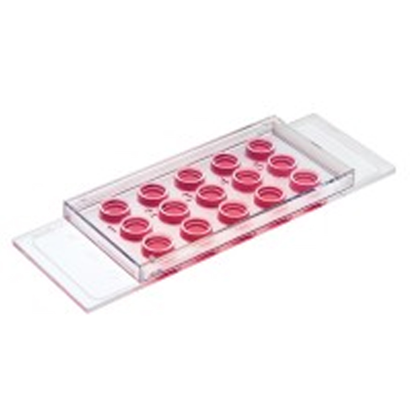
Number of wells | 8 |
Dimensions of wells (w x l x h) | 9.4 x 10.7 x 9.3 mm3 |
Volume per well | 300 μl |
Total height with/without lid | 10.8/9.5 mm |
Growth area per well | 1.0 cm2 |
Coating area per well | 2.2 cm2 |
Bottom: ibidi Polymer Coverslip | |
Technical Drawing
Technical drawings and details are available in the Instructions (PDF).
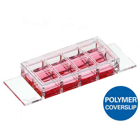
*Chambered coverslip with 8 independent wells and a non-removable polymer coverslip-bottom
*ibiTreat (tissue culture-treated) surface for optimal cell adhesion
*Imaging chamber slide with excellent optical quality for high-end microscopy
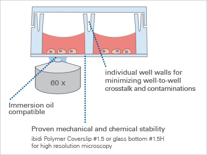
*Individual well walls for minimizing well-to-well crosstalk and contaminations
*Compatible with staining and fixation solutions
*Biocompatible polymer material—no glue, no leaking
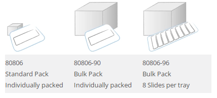
*Available as a Bulk Pack with 90 individually packed μ-Slides per box
*Also available as an adhesive version without a bottom:
sticky-Slide 8 Well high
*Also available with a Glass Coverslip Bottom:
μ-Slide 8 Well high Glass Bottom for special microscopic applications
*Additional version available with a 500 μm grid:
μ-Slide 8 Well high Grid-500
*Available with a non-adhesive Bioinert surface: μ-Slide 8 Well high Bioinert, specifically suitable for 3D applications, such as spheroids and suspension cells
*Available with a μ-Patterned surface with defined cell adhesion for single-cell and multi-cell applications

The Principle of the μ-Slide 8 Well high
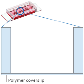
The Coverslip Bottom
The μ-Slide 8 Well high comes with a thin ibidi Polymer Coverslip Bottom that has the highest optical quality (comparable to glass) and is ideally suitable for high-resolution microscopy. It is also available as a sticky-Slide 8 Well high without any bottom and the μ-Slide 8 Well high Glass Bottom for special microscopic applications.
Find more information and technical details about the coverslip bottom of the ibidi chambers here.
The ibiTreat Surface
ibiTreat (tissue culture-treated) is our most recommended surface modification, because almost all adherent cells grow well on it without the need for any additional coating.
Find more information about the different surfaces of the ibidi chambers here.

Immunofluorescence
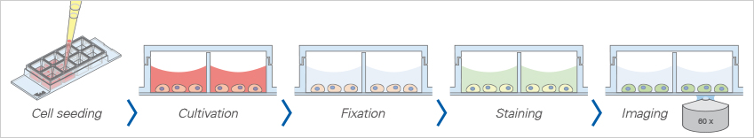
The ibidi μ-Slide 8 Well high allows for standard immunofluorescence protocols to be employed without the use of coverslips in an all-in-one chamber. All steps (e.g., cell cultivation, fixation, staining, and imaging) are carried out in the open well geometry. After staining, the sample can be observed through the coverslip bottom using high-resolution microscopy.
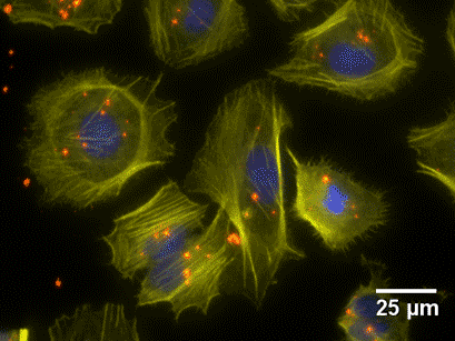
Fluorescence microscopy of HuH7 cells after mRNA transfection with eGFP (green). Actin cytoskeleton (Phalloidin, yellow), nuclei (DAPI, blue), mRNA (FISH,Quasar 670, red), 40x objective lens, oil immersion.
Live Cell Imaging
The μ-Slide 8 Well high enables high-resolution live cell imaging using different microscopy techniques.
Together with the ibidi Stage Top Incubation System, you can keep your cells happy for a long time by precisely controlling temperature, humidity, and CO2 concentration on your microscope.

Timelapse phase contrast microscopy of rat fibroblast cells using the μ-Slide 8 Well high ibiTreat in combination with the ibidi Heating System and the ibidi Gas Incubation System. 63x objective lens, oil immersion, 3 min image intervals for 23 hours.