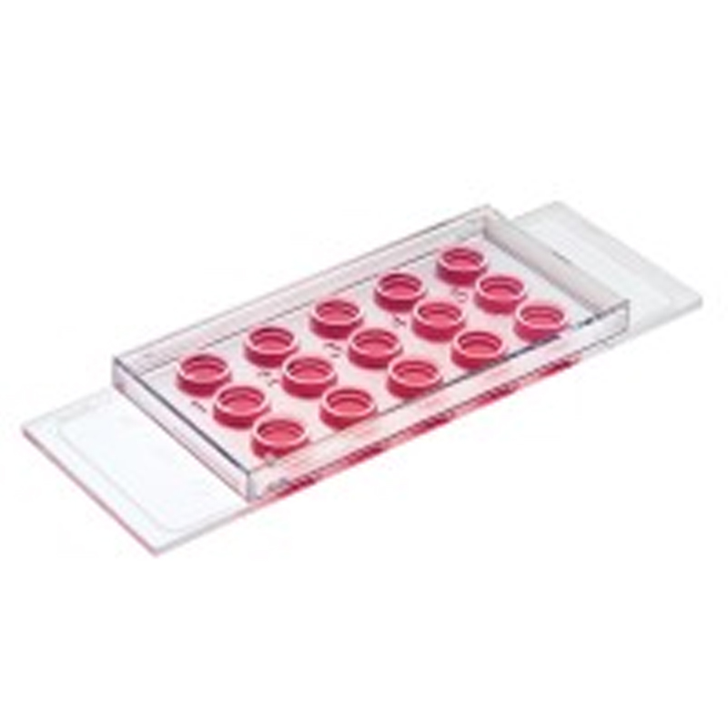

Model:81506 81501 81531
Brand:ibidi
Specifications :15 (individually packed)
Introduction:A chambered coverslip with 15 wells for 3D cell culture, tube formation assays, and immunofluorescence staining.Brilliant cell visualization without gel meniscus formation, and with all cells in one focal plane


Applications
Tube formation assays and angiogenesis assays
3D cell culture
Immunofluorescence staining
Sprouting assays
Live cell imaging of attached cells
Specifications
| Outer dimensions (w x l) | 25.5 x 75.5 mm2 |
| Number of wells | 15 |
| Volume inner well | 10 μl |
| Diameter inner well | 4 mm |
| Depth inner well | 0.8 mm |
| Volume upper well | 50 μl |
| Diameter upper well | 5 mm |
| Growth area inner well | 0.125 cm2 |
| Coating area using 10 μl | 0.23 cm2 |
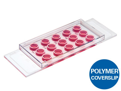
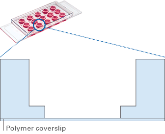
| Bottom: ibidi Polymer Coverslip | |
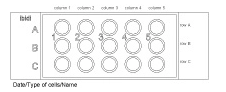
Define and print your experimental setup
Technical Features
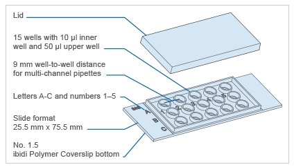
Standard slide format
Closely fitting lid to reduce evaporation
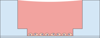
4 mm well within a 5 mm well
Geometry enables pipetting of a homogeneous 0.8 mm thick gel layer*
Provides homogeneous cell growth
Compatible with staining and fixation
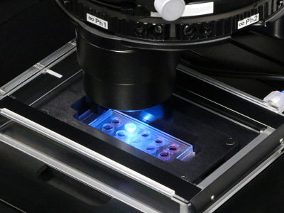
Excellent optical properties for microscopy
Compatible with multi-channel pipettes
Made of biocompatible plastic material: no glue, no leaking
Suitable for use with various types of gels* (e.g., Matrigel?, collagen, and agarose)
Also available with a No. 1.5H glass bottom: μ-Slide 15 Well 3D Glass Bottom
Also available in a 96 well format: μ-Plate 96 Well 3D
*The gel matrix is not included with this product.
Every Cell in Focus:
The Principle of the μ-Slide 15 Well 3D
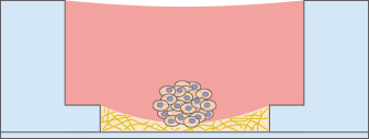
The μ-Slide 15 Well 3D has a specialized geometry for the easy, convenient, and reproducible conduction of tube formation assays. It is also ideal for sprouting assays, immunofluorescence staining, and general 3D cell culture.
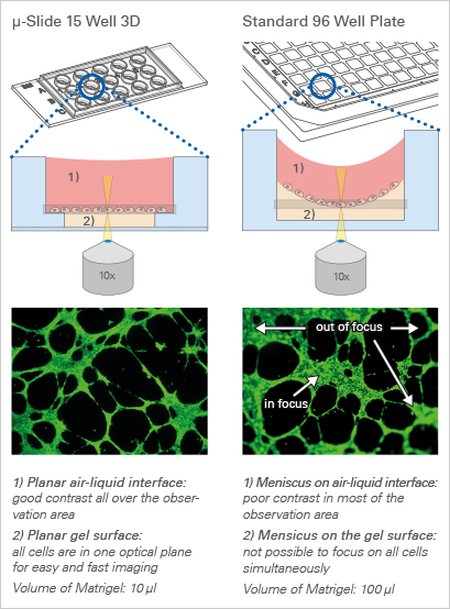
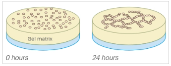
After the gel has been pipetted into the inner well and given time to solidify, the cells can be seeded on top of it for tube formation analysis. Due to the “well-in-a-well” technology, the amount of gel needed is reduced to only 10 μl per well, which is 10% of the amount used in regular multiwell plates.
Further, no gel meniscus is formed. This ensures the formation of a uniformly thick gel matrix on which all cells are in one optical plane, creating reproducible cell culture conditions. The μ-Slide 15 Well 3D can be used with all common hydrogel matrices, such as Matrigel?, collagen gels, agarose gels, and hyaluronic acid gels.
With its 15 wells, the μ-Slide 15 Well 3D is designed for low throughput assays. For large scale applications, ibidi provides the μ-Plate 96 Well 3D.
The μ-Slide 15 Well 3D Can Be Used in a Wide Range of Applications
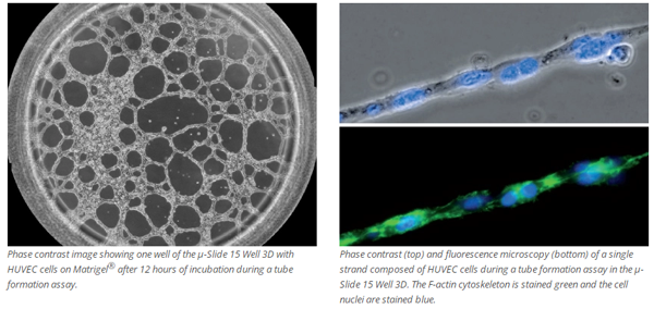
Tube Formation Assay or Angiogenesis Assay
For a tube formation assay (also called angiogenesis assay), endothelial cells are seeded on top of a 10 μl gel layer (basement membrane-like surface), where they form capillary-like structures. Typically, Matrigel? is used. The tube formation ability of the cells can be used to analyze the pro- or anti-angiogenic potential of various drugs.
Sprouting Assay
The sprouting potential of a cell cluster can be conveniently analyzed using the μ-Slide 15 Well 3D. To carry out a sprouting assay, spheroids or pieces of tissue (e.g., from the aorta), are placed onto or directly in the gel matrix before imaging.

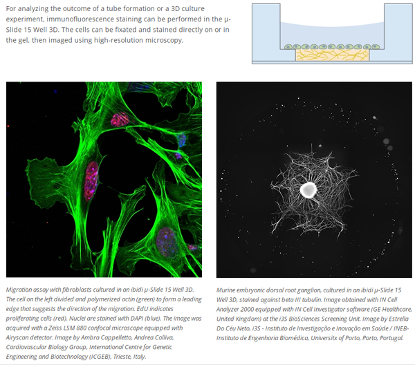
Immunofluorescence Staining
For analyzing the outcome of a tube formation or a 3D culture experiment, immunofluorescence staining can be performed in the μ-Slide 15 Well 3D. The cells can be fixated and stained directly on or in the gel, then imaged using high-resolution microscopy.
Focusing Cells
When using less than 10 μl of gel to fill the inner well of the μ-Slide 15 Well 3D, small numbers of cells can be cultivated and focused on a soft gel surface.
Low Volume Microscopy Chamber
The μ-Slide 15 Well 3D can also be used as a low-volume microscopy chamber without any gel, which is useful for experiments where several wells with a low volume are required.